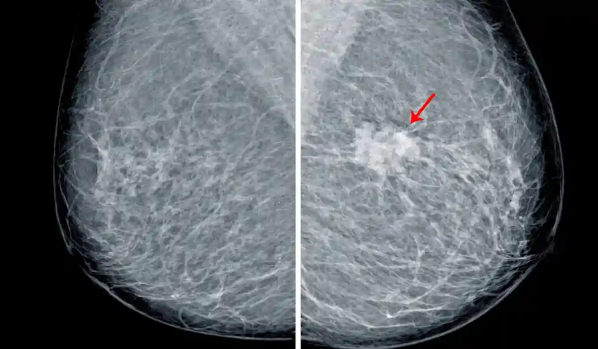
Breast calcification on mammogram means small calcium deposits were seen in breast tissue. Most breast calcifications are benign, especially larger ones, but some tiny grouped calcifications may need extra mammogram views, short-term follow-up, or biopsy to rule out early breast cancer.
Calcifications are usually too small to feel during a breast exam. A radiologist looks at their size, shape, pattern, and whether they are new or changing compared with older mammograms.
Breast Calcification on Mammogram
Breast calcifications are calcium deposits that appear as small white spots or specks on a mammogram. They are common and are often linked with aging, old injury, inflammation, cyst changes, or previous breast procedures.
The main concern is not the calcium itself. The radiologist looks at whether the calcifications are large or tiny, scattered or grouped, round or irregular, stable or new, and whether the pattern looks benign or suspicious.
Reasons for a mammogram include routine breast cancer screening, evaluation of a breast lump, nipple changes, breast pain, skin changes, or follow-up after an abnormal imaging result. A mammogram may also be used to monitor breast calcifications, compare new findings with older images, or check breast tissue after previous treatment.
What Is a Diagnostic Mammogram?
A diagnostic mammogram is a more detailed breast X-ray used when a screening mammogram shows an abnormal finding or when a person has breast symptoms. It may be recommended for a breast lump, nipple discharge, breast pain, skin changes, or breast calcifications that need closer review.
Unlike a routine screening mammogram, a diagnostic mammogram may include extra views, magnification images, or focused images of one area. Therefore, it helps the radiologist study the size, shape, and pattern of breast calcifications more clearly before deciding whether follow-up or biopsy is needed.
Breast Calcifications at a Glance
| Mammogram finding | What it usually means | Common next step |
| Macrocalcifications | Larger calcium spots, often benign | Routine screening in many cases |
| Scattered microcalcifications | Tiny specks spread out | Often follow radiologist guidance |
| Clustered microcalcifications | Tiny deposits grouped together | Diagnostic mammogram or biopsy may be needed |
| New calcifications | New compared with old images | Extra views or closer review |
| Stable benign calcifications | No concerning change over time | Routine screening or follow-up |
| BI-RADS 3 | Probably benign | Short-term imaging follow-up |
| BI-RADS 4 or 5 | Suspicious or highly suspicious | Biopsy usually recommended |
What Are Breast Calcifications?
Breast calcifications are tiny deposits of calcium in breast tissue. They are not caused by eating too much calcium and are not the same as calcium buildup in bones.
On a mammogram, these deposits appear as white dots or specks. They may be described as calcifications, microcalcifications, macrocalcifications, grouped calcifications, scattered calcifications, or suspicious calcifications.
A report may sound worrying, but many mammogram calcifications are not cancer. The radiologist’s description and BI-RADS category are more important than the word “calcification” alone.
Macrocalcifications vs Microcalcifications
Macrocalcifications are larger calcium deposits. They often look coarse, round, or clearly benign on mammogram images.
These larger calcifications are commonly related to aging, inflammation, old injury, cyst changes, or previous surgery. They usually do not suggest breast cancer when their appearance is typical.
Microcalcifications are smaller white specks. Many are benign, but certain grouped, fine, linear, branching, or irregular patterns may need closer evaluation because they can sometimes be linked with ductal carcinoma in situ, also called DCIS, or other breast changes.
When Breast Calcifications Are Usually Benign?
Benign breast calcifications often have a pattern that looks stable, scattered, round, coarse, rim-like, vascular, popcorn-like, or related to old injury or surgery.
A benign result may appear as BI-RADS 2 on the mammogram report. This means the finding is not considered cancer and routine screening is usually recommended.
Sometimes a radiologist may still compare the current mammogram with old images. Stability over time can support a benign interpretation.
When Calcifications May Be Suspicious?
Calcifications may be more concerning when they are tiny, new, tightly grouped, irregular, linear, branching, or arranged along a duct pattern.
Suspicious calcifications do not mean you definitely have cancer. They mean the appearance is concerning enough that the radiologist wants more information.
The next step may be a diagnostic mammogram with magnification views, comparison with older images, short-term follow-up, or a breast biopsy.
What BI-RADS Means for Calcifications?
BI-RADS is the standardized system radiologists use to report mammogram findings and recommended follow-up.
| BI-RADS category | Meaning | What may happen next |
| 0 | Incomplete | More imaging or old mammograms needed |
| 1 | Negative | Routine screening |
| 2 | Benign | Routine screening |
| 3 | Probably benign | Short-term follow-up imaging |
| 4 | Suspicious abnormality | Biopsy may be recommended |
| 5 | Highly suggestive of cancer | Biopsy strongly recommended |
| 6 | Known biopsy-proven cancer | Imaging used for treatment planning |
For breast calcifications on a mammogram, the BI-RADS category helps guide what you should do next. Do not rely only on the wording of the report without checking the category and recommendation.
Why Extra Mammogram Views May Be Needed?
A screening mammogram is designed to look for possible breast changes. If calcifications are unclear, the imaging center may call you back for a diagnostic mammogram.
Diagnostic views can include magnification images or spot compression images. These help the radiologist see the size, shape, and pattern of the calcifications more clearly.
A callback does not automatically mean cancer. It means the radiologist needs a closer look before giving a final assessment.
Does Breast Calcification Mean Cancer?
Most breast calcifications do not mean cancer. However, certain microcalcification patterns may be linked with early breast cancer or precancerous breast changes.
The concern is higher when calcifications are new, grouped, fine, linear, branching, or changing over time. In these cases, a biopsy may be recommended to check the breast tissue under a microscope.
Only a biopsy can confirm whether suspicious calcifications are benign, atypical, DCIS, or invasive cancer.
Breast Calcifications and DCIS
DCIS stands for ductal carcinoma in situ. It means abnormal cells are found inside the milk ducts and have not spread into nearby breast tissue.
DCIS may not cause a lump or symptoms. In some people, it is found because a mammogram shows suspicious microcalcifications.
Not all microcalcifications are DCIS. The radiologist uses the pattern, location, and BI-RADS category to decide whether biopsy is needed.
What Happens During a Biopsy for Calcifications?
If a biopsy is recommended, many calcifications are evaluated with a stereotactic breast biopsy. This uses mammogram imaging to guide a needle to the exact area.
The radiologist removes small tissue samples, and the samples are checked by a pathologist. In many cases, a tiny marker clip is placed so the area can be found again if needed.
A biopsy does not mean you have cancer. It is a way to get a definite tissue diagnosis when imaging is not enough.
What If the Biopsy Is Benign?
If the biopsy is benign and matches the imaging findings, your doctor may recommend routine screening or a short-term follow-up mammogram.
The follow-up plan depends on the pathology result, imaging appearance, BI-RADS category, and whether the radiologist and pathologist agree.
Ask whether the result is “concordant.” This means the biopsy result explains what was seen on the mammogram. If the result does not match the imaging concern, more testing may be needed.
What If the Calcifications Are Atypical or Cancerous?
If biopsy shows atypical cells, DCIS, or cancer, your care team will explain the next steps. This may include surgery consultation, additional imaging, or treatment planning.
Treatment depends on the exact diagnosis, size and location of the abnormal area, hormone receptor status when relevant, breast density, personal health, and patient preferences.
Do not assume all suspicious calcifications lead to the same treatment. The pathology report is what guides the care plan.
Symptoms to Watch For
Breast calcifications usually do not cause symptoms. Most are found only because a mammogram detects them.
Still, contact a healthcare provider if you notice:
- A new breast lump
- Nipple discharge, especially bloody discharge
- Nipple inversion that is new
- Skin dimpling or thickening
- Breast swelling or redness
- Persistent focal breast pain
- A change in breast shape
- A lump in the armpit
These symptoms may have benign causes, but they should be evaluated.
Common Mistakes to Avoid
One mistake is assuming all calcifications are cancer. Many are benign and only need routine screening or follow-up.
Another mistake is ignoring the follow-up plan. If your report recommends diagnostic mammography, biopsy, or short-term imaging, schedule it promptly.
A third mistake is comparing your report with someone else’s report. Calcification size, shape, pattern, and BI-RADS category can change the meaning.
What to Do Next After the Report?
First, read the final impression and BI-RADS category on your mammogram report. These sections usually explain the level of concern and next step.
Second, check whether the radiologist compared your new mammogram with old images. Prior images can show whether calcifications are stable or new.
Third, ask your doctor or imaging center when follow-up should happen. Keep a copy of your report and bring it to future breast imaging appointments.
Questions to Ask Your Doctor or Radiologist
- Are my breast calcifications macrocalcifications or microcalcifications?
- What BI-RADS category was assigned?
- Do the calcifications look benign, probably benign, or suspicious?
- Are they new or stable compared with old mammograms?
- Do I need magnification views or a diagnostic mammogram?
- Is ultrasound useful in my case?
- Do I need a stereotactic breast biopsy?
- If biopsy is benign, when is my next mammogram?
- What does “concordant” mean in my biopsy result?
- Should my breast density or family history change my screening plan?
Conclusion
A breast calcification on mammogram usually means calcium deposits were seen in breast tissue. Most are benign, but some microcalcification patterns need closer imaging or biopsy to rule out DCIS or breast cancer.
Overall, the safest next step is to follow the BI-RADS recommendation, compare with older mammograms when possible, and ask your healthcare provider what follow-up is needed.
FAQs
Breast calcification on mammogram means calcium deposits were seen in breast tissue. Most are benign, but certain tiny grouped or irregular patterns may need more imaging or biopsy.
Most breast calcifications are not cancer. However, suspicious microcalcifications can sometimes be linked with DCIS or breast cancer, so follow-up depends on their pattern and BI-RADS score.
Macrocalcifications are larger calcium deposits and are usually benign. Microcalcifications are smaller specks; however, grouped or irregular patterns may need closer evaluation.
A diagnostic mammogram gives magnified or special views. As a result, these images help the radiologist study the shape, size, and pattern of calcifications more clearly.
Breast calcifications usually do not cause pain and are often too small to feel. However, breast pain should be discussed with a clinician because many causes are possible.
Breast calcifications are not caused by calcium in your diet or calcium supplements. Instead, they form locally in breast tissue for reasons such as aging, inflammation, injury, or cell changes.