Prostate MRI with and without contrast helps doctors examine the prostate gland, nearby tissues, and possible abnormal areas in detail. A doctor may recommend it after a high PSA test, an unusual digital rectal exam, or before deciding whether a prostate biopsy is needed. MRI uses strong magnets and radio waves, not radiation.
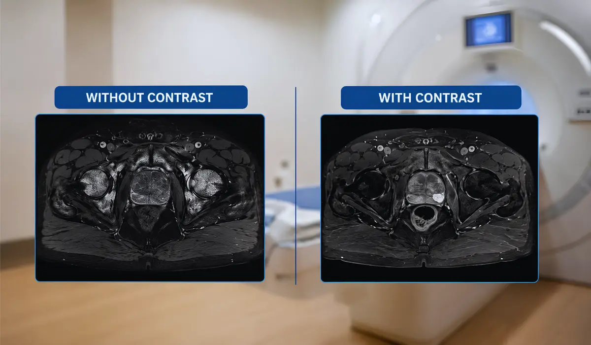
A prostate MRI without contrast uses detailed image types to study prostate size, shape, tissue pattern, and water movement. These images may help radiologists spot areas that look different from normal prostate tissue. Many noncontrast scans include anatomy images and diffusion-weighted imaging, which can show how dense or restricted tissue appears.
A prostate MRI with contrast includes an injected dye, usually gadolinium-based contrast, through a vein in the arm. This dye helps show blood flow patterns inside the prostate. Some suspicious areas absorb contrast faster or differently, which may help doctors assess unclear findings and plan biopsy or treatment decisions.
Why Doctors Recommend A Prostate MRI?
Doctors may recommend a prostate MRI when PSA results, symptoms, or a physical exam do not give enough detail. PSA can rise from prostate enlargement, inflammation, infection, recent ejaculation, or cancer. MRI helps doctors view the gland more clearly and check if any area looks suspicious.
A prostate MRI can also help guide a targeted biopsy. Instead of sampling the prostate only in a standard pattern, doctors can use MRI findings to focus on areas that appear more concerning. This can improve the accuracy of the evaluation and may help reduce unnecessary sampling in selected patients.
Doctors may also use prostate MRI after prostate cancer is diagnosed. The scan can help show whether cancer appears limited to the prostate or may have reached nearby tissues. This information supports treatment planning, including active surveillance, surgery, radiation therapy, or follow-up when PSA changes after treatment.
Key Differences Between MRI With And Without Contrast
The main difference between prostate MRI with and without contrast is the use of an injected dye. A noncontrast MRI uses detailed structural and diffusion images to show prostate anatomy and tissue changes. A contrast MRI adds images that show how prostate tissue absorbs and releases dye over time.
A prostate MRI without contrast may be shorter and does not require an IV injection. It can be useful for patients with kidney concerns, previous contrast reactions, or anxiety about needles. It may also work well when the question is simple and the imaging center uses a strong prostate MRI protocol.
A prostate MRI with contrast may be preferred when doctors need extra detail. Contrast can help assess unclear lesions, post-treatment changes, inflammation, or staging questions. Noncontrast MRI may be enough for some patients, but contrast can give radiologists more confidence when the findings are difficult to interpret.
When Prostate MRI Without Contrast May Be Enough?
A prostate MRI without contrast may be enough for some patients during an early prostate cancer check. This depends on PSA level, prostate size, symptoms, previous test results, and scan quality. If the images clearly show the prostate zones and any suspicious areas, the scan can still guide the doctor.
Noncontrast MRI may also be useful for patients who should avoid gadolinium contrast. This may include people with kidney disease, past contrast reactions, or other medical concerns. Before the scan, the imaging team may ask about kidney function, allergies, implants, medications, and previous MRI experiences.
Without contrast does not mean the scan is basic or low quality. A well-done prostate MRI without contrast can give helpful information when performed with a strong protocol and read by an experienced radiologist. However, contrast may still be preferred for cancer staging, treatment planning, or unclear previous results. Patients should ask why a specific MRI type was chosen.
When Prostate MRI With Contrast May Be Better?
Prostate MRI with contrast may be better when doctors need extra detail about a suspicious or unclear finding. Contrast helps show blood flow patterns inside the prostate. If an area looks borderline on regular MRI images, contrast may give the radiologist more information before making a final interpretation.
Doctors may also prefer contrast when prostate cancer has already been diagnosed. In this situation, the scan may help show whether cancer appears limited to the prostate or may involve nearby tissues. This information can support treatment planning for surgery, radiation therapy, or other care options.
Contrast can also help after a biopsy, procedure, or treatment, when scar tissue, inflammation, or treatment changes make the prostate harder to read. However, contrast is only one part of the MRI. The final report also depends on all image types, PSA history, physical exam findings, previous biopsy results, and the patient’s overall medical history.
How To Prepare For A Prostate MRI?
Preparation for prostate MRI can vary by imaging center, so follow the instructions from your doctor or radiology office. Many centers ask patients to avoid heavy meals before the scan. Some may suggest light bowel preparation, a mild enema, or avoiding gas-producing foods to improve image quality.
Before the MRI, tell the staff if you have a pacemaker, metal implant, artificial joint, aneurysm clip, hearing implant, or metal fragments in your body. Also mention kidney disease, allergies, past contrast reactions, claustrophobia, or trouble lying still. If contrast is planned, the team may check kidney function or recent blood test results.
On the scan day, wear clothing without metal if allowed, or change into a gown. Remove jewelry, watches, cards, keys, and other metal items before entering the MRI room. Arrive early for safety screening and paperwork. If closed spaces make you anxious, ask about calming options before the appointment, as some medicines require a ride home.
What Happens During The MRI Scan?
During a prostate MRI, you usually lie on your back on a narrow table that moves into the scanner. The machine may feel tight and make loud tapping or knocking sounds while taking images. Ear protection is usually provided, and you may get a call button to contact the technologist.
If your exam uses contrast, a small IV line is placed in your arm or hand. The contrast dye is injected during part of the scan while images are being taken. Some people notice a cool feeling, mild warmth, or a brief taste, which usually passes quickly.
The scan often takes about 30 to 60 minutes, depending on the imaging center and MRI protocol. Staying still is important because movement can blur the pictures. After the scan, most people return to normal activities. If you received sedation for anxiety, you may need monitoring and someone to drive you home. A radiologist later reviews the images and sends the report to your doctor.
Safety, Risks, And Who Should Be Careful?
Prostate MRI is generally safe for many patients because it does not use ionizing radiation. Instead, it uses a strong magnet and radio waves to create detailed images. However, the magnet can affect certain implants, devices, or metal objects, so MRI safety screening is important before the scan.
Patients should tell the imaging team about pacemakers, metal implants, aneurysm clips, hearing implants, artificial joints, or metal fragments in the body. Do not guess about past surgeries or implanted devices. If you are unsure, bring your implant card or speak with your doctor before the appointment.
For prostate MRI with contrast, doctors also review kidney function, kidney disease history, and previous contrast reactions. Gadolinium contrast is commonly used, but it may not be suitable for everyone, especially people with serious kidney disease. Allergic-type reactions are uncommon, but they can happen. If you worry about contrast or gadolinium retention, ask whether the added detail is necessary for your case. Do not cancel or change the test without medical guidance.
Choosing The Right Prostate MRI
Prostate MRI with and without contrast can both support prostate care, but the right choice depends on the reason for the scan. Doctors consider PSA results, previous tests, symptoms, and what they need to check next. A noncontrast MRI may be enough when image quality is strong and contrast is not medically needed.
Patients should know that contrast does not automatically make an MRI perfect, and a scan without contrast is not automatically weak. The scanner quality, MRI protocol, and radiologist’s prostate imaging experience all matter. PSA trend, age, prostate size, family history, symptoms, and biopsy history also affect how results are understood.
Before the appointment, ask your doctor why the MRI is needed, whether contrast will be used, and what the scan is meant to rule out or confirm. Also ask what may happen after the report is ready. Prostate MRI does not diagnose everything by itself, but it can help doctors make clearer, more personalized decisions.
FAQs
It can be better in some cases because contrast adds blood-flow information. However, noncontrast MRI may still work well for selected patients.
It may miss some findings, like any imaging test. Accuracy depends on scan quality, radiologist experience, PSA pattern, and whether biopsy is also needed.
Doctors may avoid contrast if you have kidney problems, a past contrast reaction, low-risk evaluation needs, or a situation where noncontrast images are enough.
Not always. Your doctor reviews the MRI report with PSA, exam findings, symptoms, age, and risk factors before deciding if biopsy is needed.
Follow your imaging center’s instructions, share kidney and allergy history, remove metal items, arrive early, and ask about anxiety medicine if needed.
References
Urology Times
Expert Discusses Use of Prostate MRI With and Without Contrast
https://www.urologytimes.com/view/expert-discusses-use-prostate-mri-with-and-without-contrast
RadiologyInfo.org
Prostate MRI
https://www.radiologyinfo.org/en/info/mr_prostate
Mayo Clinic
Prostate MRI
https://www.mayoclinic.org/tests-procedures/prostate-mri/about/pac-20596560
American College of Radiology
Prostate Imaging Reporting & Data System — PI-RADS
https://www.acr.org/Clinical-Resources/Clinical-Tools-and-Reference/Reporting-and-Data-Systems/PI-RADS