A prostate MRI is a detailed imaging test that helps doctors examine the prostate more clearly. It can support decisions about biopsy, monitoring, diagnosis, and treatment planning.
What Is a Prostate MRI?
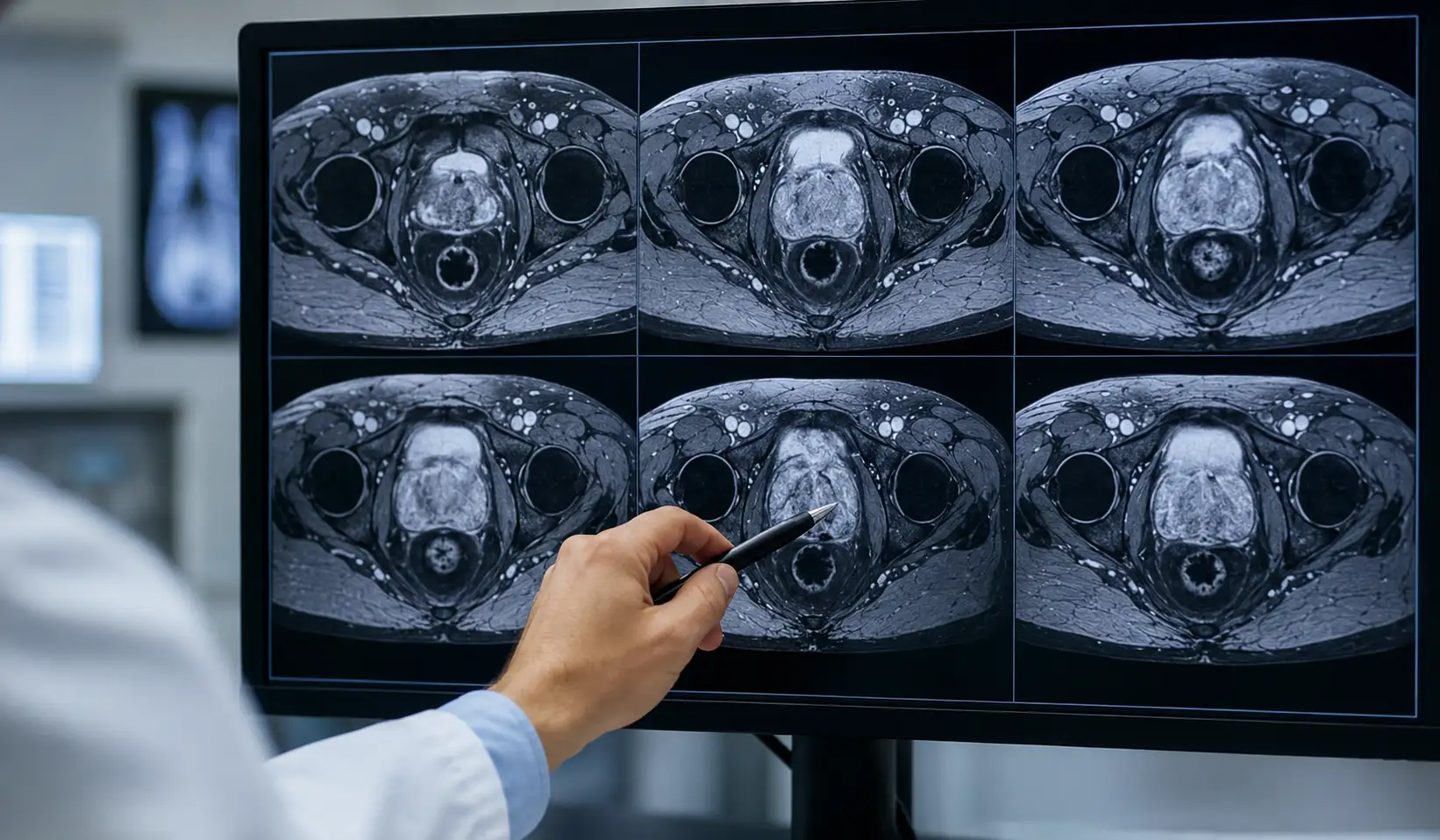
A prostate MRI is an imaging test that creates detailed pictures of the prostate gland. Doctors use it to look more closely at prostate tissue when a PSA blood test, physical exam, symptoms, or previous test results need more explanation.
MRI stands for magnetic resonance imaging. The test uses a strong magnetic field and radio waves to build images of the inside of the body. It does not use radiation, which makes it different from X-rays and many CT scans.
A prostate MRI does not replace every other prostate test. Instead, it gives doctors extra information. It may help find suspicious areas, guide a biopsy, review prostate cancer after diagnosis, or check non-cancer prostate conditions such as enlargement or inflammation.
Why Doctors Recommend A Prostate MRI?
Doctors may recommend a prostate MRI after a high PSA test, an abnormal digital rectal exam, or ongoing urinary or pelvic symptoms. The scan can show details that basic testing cannot always provide, so it may help doctors decide what should happen next.
One common reason is biopsy planning. If the MRI scan shows an area that looks concerning, the doctor may target that spot during a prostate biopsy. This approach can help focus testing on the area most likely to need attention.
After prostate cancer is diagnosed, MRI may help doctors understand whether the cancer appears limited to the prostate or has grown nearby. It may also support active surveillance, where doctors monitor low-risk cancer closely instead of treating it right away.
How To Prepare For A Prostate MRI?
Preparation can vary by imaging center, so follow the instructions from your care team. Some people may be asked to stop eating or drinking for a short period before the scan. Others may continue their normal routine unless told otherwise.
Tell your healthcare team about all medicines, vitamins & supplements, allergies, kidney disease, and past reactions to contrast material. Also mention implanted devices, pacemakers, glucose monitors, hearing implants, metal clips, bullet fragments, piercings, or any metal that cannot be removed.
Some prostate MRI scans use contrast through a vein to improve detail. A few exams may use a small rectal device called an endorectal coil. If this is planned, you may receive bowel preparation instructions, such as using an enema before the appointment.
What Happens During The Scan?
Before the scan, you usually change into a gown and remove metal items such as jewelry, watches, piercings, hearing aids, and removable dental devices. The team may also ask you to use the bathroom because an empty bladder or rectum can improve image quality.
During the prostate MRI, you lie still on a table that slides into the MRI machine. The machine often looks like a long tube open on both ends. The technologist moves to another room, but the team can see, hear, and speak with you.
The test should not be painful, but staying still may feel uncomfortable. MRI machines make tapping or thumping sounds, so you may receive earplugs or music. Many prostate MRI scans take about 45 minutes when contrast is used, while non-contrast scans may take less time.
Safety, Comfort, And Possible Risks
A prostate MRI is generally safe for most people. It does not use radiation, and standard MRI magnetic fields and radio waves are not known to cause harmful effects when proper screening rules are followed.
The biggest safety concern involves metal. Strong magnets can affect some implanted devices or metal objects and may also blur images. Your care team screens for these risks before the scan, so give complete and honest information about anything metal in your body.
Contrast material is safe for many people, but side effects can happen. Some people get bruising at the injection site, nausea, or rarely an allergic reaction. People with kidney disease may need kidney function testing before certain gadolinium contrast agents are used.
Understanding Prostate MRI Results And Next Steps
A radiologist reviews the prostate MRI images and writes a report for the doctor who ordered the test. The imaging staff usually cannot explain results during the appointment. Your doctor may call you, schedule a visit, or release the report through a patient portal.
The report may use terms such as lesion, restricted diffusion, low T2 signal, DCE positive, or PI-RADS score. A PI-RADS score estimates how likely a suspicious area is to be clinically significant prostate cancer, usually on a scale from 1 to 5.
A prostate MRI can help guide decisions, but it cannot prove cancer by itself. It can miss cancer or show changes caused by inflammation, enlargement, or bleeding. Your doctor will review MRI findings with PSA results, symptoms, exam findings, and biopsy results before recommending next steps.
FAQs
A prostate MRI is not usually painful. You may feel discomfort from lying still, machine noise, contrast injection, or rectal pressure if a coil is used.
A prostate MRI can show suspicious areas and estimate cancer likelihood, but a biopsy is usually needed to confirm prostate cancer with tissue testing afterward.
A prostate MRI often takes about 45 minutes when contrast is used. Non-contrast scans may take less time depending on your imaging center’s protocol.
Not always. Some prostate MRI exams use gadolinium contrast to show blood flow, while others rely on non-contrast techniques chosen by your doctor carefully beforehand.
Ask about your PI-RADS score, whether biopsy is needed, what findings mean with your PSA level, and when follow-up testing should happen next with your doctor.
References
Mayo Clinic – Prostate MRI
https://www.mayoclinic.org/tests-procedures/prostate-mri/about/pac-20596560
Johns Hopkins Medicine – Radiology Exam: Prostate MRI
https://www.hopkinsmedicine.org/radiology/specialties/prostate-mri
RadiologyInfo.org – Prostate MRI
https://www.radiologyinfo.org/en/info/mr_prostate
National Cancer Institute – Prostate Cancer Screening
https://www.cancer.gov/types/prostate/hp/prostate-screening-pdq
RadiologyInfo.org – How to Read Your Prostate MRI Report
https://www.radiologyinfo.org/en/info/article-prostate-mri-report