Nerve pain can be confusing because the problem is not always easy to see from the outside. A person may feel burning, tingling, numbness, weakness, or sharp pain, but the exact cause may come from the spine, brain, muscles, bones, or the nerve itself.
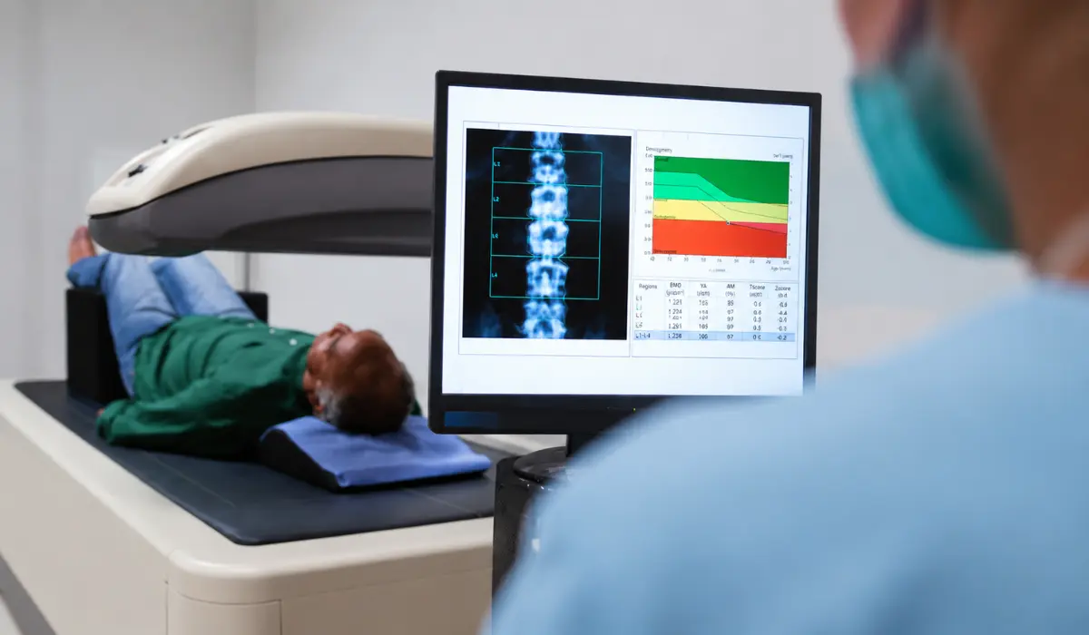
When symptoms continue, doctors may suggest imaging tests. The common question is whether an MRI or CT scan for nerve damage is better. In many cases, MRI is preferred for soft tissues and nerves, while CT is more useful for bones, fractures, emergency injuries, and certain spine problems. MRI also provides better soft tissue contrast than CT, according to the FDA.
Why Doctors Use Scans For Nerve Symptoms?
Doctors do not always order a scan right away for nerve symptoms. First, they may check your pain location, muscle strength, reflexes, walking pattern, medical history, and whether symptoms started suddenly or slowly.
A scan may be recommended if symptoms are severe, getting worse, linked to injury, or not improving with basic treatment. Imaging can help find problems like a herniated disc, spinal narrowing, tumor, inflammation, fracture, or pressure on a nerve.
MRI Or CT Scan For Nerve Damage: The Main Difference
MRI is usually better when the doctor needs to see soft tissues. This includes the spinal cord, discs, muscles, ligaments, inflammation, and some nerve-related changes. Cleveland Clinic notes that MRI can show some nerves and may help show injury or inflammation in certain body areas.
CT scans are different because they are often better for bone detail and fast emergency imaging. A CT scan may not show nerves as clearly as MRI, but it can show fractures, bone spurs, spinal narrowing, or other hard-tissue problems that may press on nerves.
When MRI May Be The Better Choice?
MRI is often selected when symptoms suggest nerve compression, disc problems, spinal cord issues, or soft tissue injury. For example, if someone has back pain with leg numbness, neck pain with arm weakness, or suspected pinched nerves, MRI may provide more helpful detail.
MRI may also be useful when doctors need to check inflammation, tumors, or changes around the spinal cord. RadiologyInfo says spine MRI is used to help plan procedures for pinched nerves, spinal fusion, or steroid injections.
When CT Scan May Be The Better Choice?
A CT scan may be chosen when a doctor needs a fast look at bones, bleeding, trauma, or fractures. It is commonly used in emergency settings because it is quick and widely available.
CT may also be used when a person cannot safely have an MRI. For example, some people with certain implanted devices, metal fragments, or severe claustrophobia may not be ideal MRI candidates. Johns Hopkins Medicine explains that CT may be recommended when a patient cannot have an MRI.
Can A Scan Directly Show Nerve Damage?
A scan may show the reason for nerve symptoms, but it may not always prove how well the nerve is working. For example, MRI can show a herniated disc pressing on a nerve, but the doctor may still need other tests to measure nerve function.
This is why doctors may also suggest nerve conduction studies or EMG testing. These tests check how electrical signals move through nerves and muscles. Imaging shows structure, while nerve tests can help show function.
MRI Neurography For Nerve Pain
In some cases, a regular MRI may not be enough. Doctors may suggest MR neurography, which is a special type of MRI designed to view peripheral nerves in more detail.
Hospital for Special Surgery explains that MR neurography uses high-resolution imaging to show nerves throughout the body and may help detect abnormal nerve size, brightness, injury, or inflammation.
Which Scan Is Safer?
MRI does not use ionizing radiation, which is one reason it is often preferred when soft tissue detail is needed. However, MRI uses a strong magnetic field, so safety screening is important before the scan.
CT scans use X-rays, so they involve radiation exposure. This does not mean CT is unsafe, but doctors usually order it when the benefit is greater than the risk. The best scan depends on the patient’s condition, urgency, and medical history.
Symptoms That May Need Imaging
Nerve symptoms can happen for many reasons. Common symptoms include tingling, numbness, burning pain, weakness, shooting pain, balance problems, or pain that travels from the neck to the arm or from the lower back to the leg.
Medical attention is more urgent if symptoms appear suddenly, affect one side of the body, cause loss of bladder or bowel control, follow an injury, or come with severe weakness. These signs may need faster testing and treatment.
Final Thoughts
For most nerve-related concerns, MRI is often the better imaging test because it gives clearer detail of soft tissues, discs, the spinal cord, and some nerve-related changes. It is commonly used when doctors suspect nerve compression, inflammation, or spine-related causes.
A CT scan still has an important role. It may be better for fractures, bone problems, trauma, and emergency situations. The right choice depends on your symptoms, health history, and what your doctor is trying to confirm.
FAQ
No, MRI is usually better for nerve-related problems because it shows soft tissues, discs, inflammation, and nerve compression more clearly than a CT scan can.
Doctors may prefer CT in emergencies because it is faster, widely available, better for bones and trauma, and easier for patients who cannot stay still.
A standard CT usually cannot show nerve damage directly, but it may reveal bone spurs, fractures, or disc issues pressing on a nerve root nearby.
Some nerve damage may improve with treatment, time, therapy, or surgery, but recovery depends on the cause, severity, location, and how quickly care starts afterward.
MRI is safer regarding radiation because it uses none, while CT uses X-rays; however, both are considered safe when medically needed and properly ordered carefully.