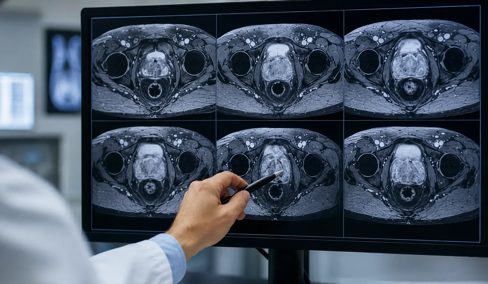
A normal vs abnormal PET scan can help doctors understand how active certain cells and tissues are inside the body. A normal PET scan usually shows expected tracer activity, while an abnormal PET scan may show areas with unusual uptake that need closer review.
PET scans do not diagnose every condition by themselves. Doctors compare the scan with symptoms, medical history, blood tests, biopsy results, CT scans, MRI scans, and previous imaging before making a final decision.
What Is Normal vs Abnormal PET Scan?
A PET scan, or positron emission tomography scan, uses a small amount of radioactive tracer to show how tissues use energy. Many PET scans use FDG, a sugar-like tracer that collects in cells with higher activity.
A normal PET scan shows tracer activity in expected places, such as the brain, heart, liver, kidneys, bladder, and some muscles. These areas naturally use energy, so uptake does not always mean disease.
An abnormal PET scan shows unusual tracer uptake, low activity, or a pattern that does not fit normal body function. This may point to cancer, inflammation, infection, heart problems, or brain disorders, depending on the scan type.
Why Normal vs Abnormal PET Scan Matters?
People search for normal vs abnormal PET scan because PET results can feel confusing. A report may mention “uptake,” “FDG avid,” “SUV,” “lesion,” or “metabolic activity,” and these words can sound worrying.
Understanding the basic difference can reduce fear and help patients ask better questions. Still, only a radiologist and treating doctor can explain what the result means for one person.
PET scans matter because they show function, not just structure. CT and MRI show body anatomy in detail, while PET shows how active tissues are. This can help doctors find disease, check spread, or monitor treatment response.
What a Normal PET Scan May Show?
A normal PET scan does not mean the whole body has zero tracer uptake. Some organs naturally look active because they use more energy.
The brain often shows strong activity because it uses glucose. The heart may also show uptake, depending on diet, fasting, and scan purpose. Kidneys and bladder commonly appear bright because the body removes the tracer through urine.
Muscles can show uptake if a person moved, exercised, shivered, or talked too much before the scan. Brown fat can also appear active, especially in cold conditions.
What an Abnormal PET Scan May Show?
An abnormal PET scan may show a bright spot, unusual pattern, or unexpected tracer uptake. Doctors often call this “increased metabolic activity” or “FDG avid uptake.”
Cancer can appear abnormal because many cancer cells use more glucose than normal cells. However, inflammation and infection can also cause increased uptake. This is why a bright spot does not always mean cancer.
Some abnormal results may show reduced activity instead of increased uptake. In brain or heart imaging, low activity in certain areas may suggest reduced function, poor blood flow, or tissue damage.
Common Reasons for Abnormal PET Scan Findings
Cancer Evaluation
Doctors often use PET scans to help detect cancer, stage cancer, check whether it has spread, and monitor treatment response. A PET scan may show areas where cancer cells appear more metabolically active.
However, PET findings need confirmation. A doctor may recommend biopsy, follow-up imaging, lab tests, or comparison with older scans.
Infection or Inflammation
Infection and inflammation can also create abnormal PET activity. The immune system uses energy during healing, so inflamed tissue may look bright.
Examples may include lung infection, inflammatory bowel disease, arthritis, healing surgical sites, or recent radiation treatment. Timing and clinical history help doctors avoid misreading these changes.
Heart Conditions
Cardiac PET scans can show blood flow and heart muscle activity. Doctors may use them to check coronary artery disease, damaged heart tissue, or whether heart muscle may recover after treatment.
A normal cardiac PET scan may show good blood flow. An abnormal result may suggest reduced blood supply or damaged heart muscle.
Brain Disorders
Brain PET scans may help evaluate certain neurological conditions. Doctors may use them for dementia evaluation, seizures, or brain tumors depending on the tracer and clinical need.
Abnormal brain PET patterns do not give a final answer alone. A neurologist usually reviews the scan with symptoms, memory testing, MRI, and other exams.
Normal vs Abnormal PET Scan Comparison Table
| Feature | Normal PET Scan | Abnormal PET Scan |
| Tracer activity | Appears in expected body areas | Appears in unusual areas or patterns |
| Bright spots | May occur in organs that naturally use energy | May suggest cancer, infection, or inflammation |
| Report wording | “No abnormal uptake” or “physiologic uptake” | “FDG avid,” “hypermetabolic,” or “suspicious uptake” |
| Meaning | No concerning activity seen on that scan | Needs doctor review and possible follow-up |
| Next step | Routine follow-up if needed | More testing, biopsy, or repeat imaging may follow |
PET Scan Terms Patients Often See
FDG Uptake
FDG uptake means the tracer collected in a tissue. Some FDG uptake is normal, while unusual uptake may need more review.
SUV
SUV stands for standardized uptake value. It gives a number that estimates how much tracer a specific area takes up.
A higher SUV can suggest more activity, but it does not prove cancer by itself. Doctors look at the pattern, location, size, history, and comparison scans.
Physiologic Uptake
Physiologic uptake means normal tracer activity. It often appears in organs that naturally use energy or remove the tracer.
Hypermetabolic Activity
Hypermetabolic activity means an area uses more energy than expected. Cancer, infection, inflammation, and healing tissue can all cause this finding.
How to Understand a PET Scan Report?
Start by reading the “Impression” section. This part gives the radiologist’s summary and usually explains the most important findings.
Next, look for whether the report says the uptake is suspicious, nonspecific, inflammatory, stable, improved, or resolved. These words help explain how concerning the finding may be.
Compare the report with previous scans if available. A spot that shrinks or becomes less active after treatment may mean something different from a new or growing spot.
Things That Can Affect PET Scan Results
Several factors can change PET scan appearance. Eating before the scan, high blood sugar, recent exercise, infection, inflammation, recent surgery, and certain treatments may affect tracer uptake.
Diabetes can also affect scan preparation and image quality. Patients with diabetes should follow the imaging center’s instructions carefully.
Movement during the scan can blur images. Wearing metal items or not following preparation instructions may also affect results.
When an Abnormal PET Scan Is Not Cancer?
An abnormal PET scan does not always mean cancer. Many non-cancer problems can create bright areas.
Inflammation, infection, healing wounds, recent surgery, arthritis, radiation changes, and some benign tumors may show increased uptake. This can lead to false-positive results.
A doctor may recommend another scan, lab test, biopsy, or short-term follow-up to understand the finding better.
Can a Normal PET Scan Miss Disease?
A normal PET scan can still miss some diseases. Very small tumors, slow-growing cancers, low-grade tumors, or certain cancer types may not show strong tracer uptake.
Blood sugar levels, timing, scanner quality, and body movement can also affect image results. No imaging test gives perfect answers.
If symptoms continue despite a normal PET scan, the doctor may order additional tests. A normal result should not replace medical follow-up when symptoms remain unexplained.
Safety Notes Before and After a PET Scan
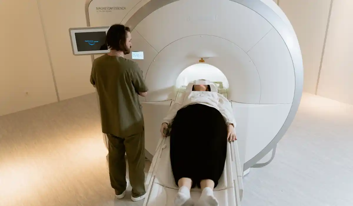
A PET scan uses a small amount of radiation. For most adults, the risk is low, but pregnant or breastfeeding patients should tell the imaging team before the scan.
Patients should follow fasting instructions before the appointment. Many centers ask patients to avoid food for several hours and avoid strenuous exercise before the scan.
After the scan, drinking water may help the body remove the tracer through urine. Follow the imaging center’s instructions, especially if you care for infants, are breastfeeding, or have kidney concerns.
When to Ask a Doctor?
Ask your doctor to explain any unclear words in your PET report. Important terms include FDG avid, suspicious uptake, lesion, lymph node activity, SUV, and metastasis.
Call your doctor sooner if you have new symptoms, worsening pain, fever, unexplained weight loss, breathing trouble, or sudden changes in health.
Do not assume the worst from one sentence in the report. PET scan results need full medical context before they can guide decisions.
Final Thoughts
Normal vs abnormal PET scan results depend on where tracer activity appears, how strong it looks, and whether the pattern fits normal body function. A normal scan usually shows expected uptake, while an abnormal scan shows unusual activity that needs review.
Bright spots can suggest cancer, but they can also come from infection, inflammation, healing, or normal organ activity. A normal scan can also miss some disease, especially if the finding is small or slow-growing.
The best next step is to review the report with your doctor. They can explain what the PET scan means in your case and whether you need follow-up imaging, lab tests, biopsy, or treatment.
FAQs
Normal vs abnormal PET scan means expected tracer activity compared with unusual uptake. Abnormal findings may need more testing but do not always mean cancer.
No. Infection, inflammation, healing tissue, recent surgery, and some benign conditions can also cause abnormal uptake on a PET scan.
A normal PET scan shows expected tracer uptake in areas such as the brain, heart, liver, kidneys, bladder, and other normal tissues.
Yes. A PET scan may miss very small, slow-growing, or low-activity cancers. Doctors may order more tests if symptoms or risks continue.
Ask what the uptake may mean, whether it looks suspicious, whether previous scans changed, and if you need follow-up imaging, biopsy, or treatment.
References
- National Cancer Institute – PET Scan Definition
https://www.cancer.gov/publications/dictionaries/cancer-terms/def/pet-scan - Mayo Clinic – PET Scan Overview
https://www.mayoclinic.org/tests-procedures/pet-scan/about/pac-20385078