A saline infusion sonogram is a special ultrasound test that gives doctors a clearer look inside the uterus. It is often used when a regular pelvic ultrasound does not show enough detail or when symptoms suggest a problem in the uterine cavity.
This test is also called saline infusion sonography, SIS ultrasound, sonohysterography, or a sonohysterogram. Doctors may recommend it for abnormal bleeding, fertility testing, suspected polyps, fibroids, scar tissue, or changes in the uterine lining.
What This Uterine Ultrasound Test Means?
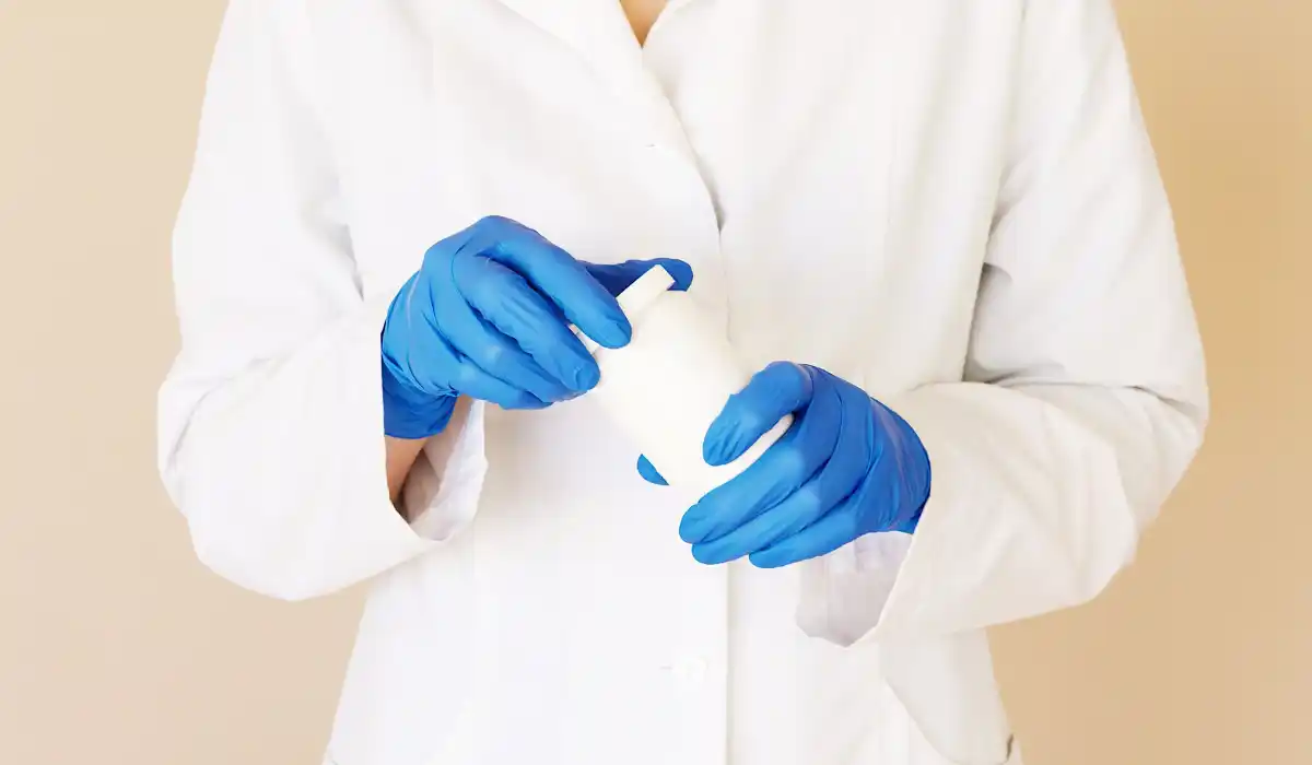
A saline infusion sonogram uses sterile salt water and ultrasound imaging to examine the inside of the uterus. During the procedure, a small amount of saline is placed through the cervix into the uterine cavity.
The fluid gently separates the uterine walls. This makes the lining easier to see on the ultrasound screen. Without saline, the front and back walls of the uterus may touch, making small growths or changes harder to identify.
The test does not use radiation. It works with sound waves, much like a standard transvaginal ultrasound. The added saline gives the doctor a more detailed view.
Why Doctors Recommend It?
Doctors often order this test when a patient has symptoms that may come from the inside of the uterus. Common reasons include heavy periods, irregular bleeding, spotting between periods, bleeding after menopause, or unclear ultrasound findings.
A saline infusion sonogram may also be used during fertility evaluation. The uterine cavity needs to be healthy for implantation and pregnancy. If there are polyps, fibroids, scar tissue, or shape changes, they may affect fertility treatment planning.
Some doctors use SIS ultrasound before IVF, IUI, or other fertility care. It can help confirm whether the uterine cavity looks normal before treatment begins.
Conditions It May Help Detect
This test can help identify several uterine problems. One common finding is an endometrial polyp, which is a small growth from the lining of the uterus. Polyps may cause spotting, heavy bleeding, or fertility concerns.
It may also show submucosal fibroids. These are fibroids that grow into the uterine cavity. Depending on size and location, they can affect bleeding patterns or pregnancy chances.
Other possible findings include uterine scar tissue, adhesions, congenital shape differences, thickened lining, or uneven areas in the endometrium. The results help your doctor decide whether monitoring, biopsy, hysteroscopy, or treatment is needed.
How the Procedure Is Done?
A saline infusion sonogram is usually done in a clinic, imaging center, or fertility office. You lie on an exam table in a position similar to a pelvic exam.
A speculum may be placed in the vagina so the cervix can be seen. The provider then passes a very thin tube through the cervix into the uterus. After that, the speculum is usually removed, and a transvaginal ultrasound probe is placed.
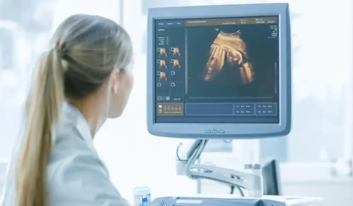
Sterile saline is slowly pushed through the tube while ultrasound images are taken. As the fluid fills the uterine cavity, the doctor can see the shape, lining, and any visible abnormal areas more clearly.
Best Time in the Menstrual Cycle
The timing of the test matters. In many cases, it is scheduled after your period ends but before ovulation. This helps reduce the chance of doing the procedure during early pregnancy and gives a clearer view of the lining.
For people with regular cycles, this may be around days 6 to 11 of the cycle. Timing can vary based on bleeding pattern, fertility treatment schedule, or medical history.
If you have irregular bleeding or do not have regular periods, your doctor may choose the safest and most useful timing for your situation.
Does a Sonohysterogram Hurt?
Many people feel mild to moderate cramping during the test. The cramps may feel similar to period pain. Some people only feel pressure, while others feel stronger discomfort when the saline enters the uterus.
The discomfort usually lasts only a short time. It often improves once the procedure is finished. Your clinic may suggest taking an over-the-counter pain reliever before the appointment, but only take medicine that is safe for you.
Tell the provider during the test if the pain feels severe. They may slow down, pause, or stop if needed.
How to Prepare Before the Appointment?
Preparation is usually simple. Your clinic may ask you to avoid sex or confirm that you are not pregnant before the test. You may also be asked to empty your bladder before the procedure.
Wear comfortable clothing and consider bringing a pad. Mild spotting or watery discharge can happen afterward because some saline may drain out.
Tell your doctor before the visit if you have fever, pelvic pain, unusual discharge, possible pregnancy, or a recent pelvic infection. The test may need to be delayed if infection is suspected.
What to Expect Afterward?
After a saline infusion sonogram, you may have light cramping, spotting, or watery discharge. These symptoms are usually mild and often improve the same day.
Most people can return to normal activities soon after the appointment. However, follow the instructions given by your healthcare provider, especially if you are having fertility treatment or another procedure soon.
Call your doctor if you develop fever, worsening pelvic pain, heavy bleeding, dizziness, chills, or foul-smelling discharge. These symptoms are not expected and should be checked.
Understanding Your Results
Some results may be explained right after the test. In other cases, the images are reviewed first, and your doctor discusses the findings later.
A normal result usually means the uterine cavity looks smooth and no clear growths or abnormal areas were seen. An abnormal result may show a polyp, fibroid, scar tissue, thickened lining, or a shape difference.
Your next step depends on the finding. Some issues may only need monitoring. Others may require biopsy, medication, hysteroscopy, or removal of a polyp or fibroid.
Regular Ultrasound vs SIS
A regular transvaginal ultrasound can show the uterus, ovaries, and nearby pelvic structures. It is often the first imaging test used for pelvic pain, bleeding, or fertility concerns.
SIS ultrasound gives a closer view of the uterine cavity because the saline outlines the lining. This makes it easier to see small growths that may not be obvious on a standard scan.
In simple terms, a regular ultrasound gives a broad pelvic view. A saline infusion sonogram gives a more detailed look inside the uterus.
SIS vs Hysteroscopy
SIS is an imaging test. It helps doctors see possible problems, but it does not treat them. Hysteroscopy uses a small camera placed inside the uterus and may allow the doctor to treat certain problems during the same procedure.
Your provider may recommend SIS first because it is less invasive and useful for diagnosis. If the test shows a polyp, fibroid, or scar tissue, hysteroscopy may be the next step.
The right choice depends on symptoms, age, fertility goals, test results, and whether treatment is likely needed.
Safety and Possible Side Effects
A saline infusion sonogram is generally considered safe for many patients, but it is not right for everyone. It is usually not done during pregnancy or when there is an active pelvic infection.
Common side effects include temporary cramps, light spotting, and watery discharge. Rare problems may include infection or stronger pain.
Before the test, tell your provider about allergies, pregnancy possibility, pelvic infection history, or current symptoms. This helps the clinic decide whether the procedure is appropriate.
When to Call Your Doctor?
Call your healthcare provider if you have heavy bleeding, fever, chills, severe pelvic pain, dizziness, or discharge with a bad smell after the test.
You should also ask for clarification if you do not understand the result. A report may include terms like polyp, fibroid, adhesion, endometrium, or uterine cavity. Your doctor can explain what the finding means for your health or fertility plan.
Final Thoughts on Saline Infusion Sonogram
A saline infusion sonogram is a helpful test for checking the inside of the uterus with more detail than a standard ultrasound. It may be recommended for abnormal bleeding, fertility testing, suspected polyps, fibroids, or unclear pelvic ultrasound results.
The procedure is usually short, does not use radiation, and often allows people to return to normal activities the same day. If your doctor recommends it, ask why it is needed, how to prepare, when results will be available, and what the next step may be.
FAQs
Your doctor may order it to check abnormal bleeding, fertility concerns, suspected polyps, fibroids, scar tissue, or unclear findings from a regular ultrasound.
The imaging part is usually short, often around 15 to 30 minutes. Total visit time may vary depending on clinic workflow.
Most people can drive home after the test. If you feel strong cramps, dizziness, or discomfort, rest first and follow clinic advice.
This test is usually not done during pregnancy. Tell your provider if you may be pregnant before the procedure is scheduled.
The main purpose is to view the uterine cavity. However, the ultrasound may also show the ovaries and other pelvic structures.
Your doctor may recommend monitoring, medication, biopsy, hysteroscopy, or removal of a polyp or fibroid depending on the result and symptoms.
References
- Cleveland Clinic – Sonohysterogram
https://my.clevelandclinic.org/health/diagnostics/22320-sonohysterogram - PMC – Utility of Saline Infusion Sonohysterography in Gynecology
https://pmc.ncbi.nlm.nih.gov/articles/PMC10040238/