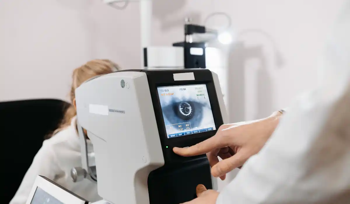
Fluorescein angiography is an eye imaging test that helps doctors see the blood vessels in the retina. The test uses a yellow dye called fluorescein and a special camera to take pictures as the dye moves through the eye’s blood vessels.
Eye doctors may recommend this retinal dye test when they need to check for leaking, blocked, or abnormal vessels. It can help diagnose diabetic retinopathy, macular degeneration, retinal vein occlusion, macular edema, and other retina problems.
What Is Fluorescein Angiography?
Fluorescein angiography is a diagnostic eye test used to examine blood flow in the retina and sometimes the choroid, which is a layer of blood vessels under the retina. It is also called FA, fluorescein angiogram, or retinal angiography.
During the test, a healthcare professional injects fluorescein dye into a vein, usually in the arm or hand. As the dye travels through the bloodstream and reaches the eye, a special camera takes a series of photographs. These images help the eye doctor study blood vessel patterns and detect problem areas.
Why Eye Doctors Use This Retinal Dye Test?
Doctors use FA test to get a clearer view of retinal blood vessels. The test can show leaking vessels, blocked blood flow, abnormal vessel growth, swelling, and areas with poor circulation.
Your eye doctor may order this test if an eye exam, vision symptoms, or another imaging test suggests a retina problem. Fluorescein angiography can help confirm a diagnosis, guide treatment, and monitor how well treatment is working.
FA Test for Diabetic Retinopathy
Diabetic retinopathy is one of the most common reasons for fluorescein angiography. High blood sugar can damage small blood vessels in the retina over time. These vessels may leak fluid, bleed, close off, or grow abnormally.
A fluorescein angiogram can help the doctor see which blood vessels are leaking or blocked. This information may help guide treatment for diabetic macular edema, severe diabetic retinopathy, or abnormal new blood vessel growth.
Fluorescein Angiogram for Macular Degeneration
Fluorescein angiography may also help diagnose and monitor wet age-related macular degeneration. Wet AMD can happen when abnormal blood vessels grow under the retina and leak fluid or blood.
The test helps the eye doctor see whether abnormal leaking vessels are present. It may also help decide whether treatment, such as eye injections, may be needed. Early diagnosis and follow-up can help protect central vision.
Other Eye Conditions Checked With Retina Imaging
Eye angiogram can help evaluate many retinal conditions. These may include retinal vein occlusion, retinal artery occlusion, macular edema, retinal inflammation, uveitis, eye tumors, and abnormal blood vessel growth.
The test may also help doctors study unexplained vision loss, swelling in the retina, or changes seen during a dilated eye exam. It is especially useful when blood vessel leakage or poor blood flow may be involved.
Fluorescein Angiography vs OCT Scan
Fluorescein angiography and OCT are both important retina tests, but they show different information. OCT creates cross-section images of the retina and helps show swelling, fluid, and retinal thickness.
Fluorescein angiography shows how dye moves through the retinal blood vessels. It can reveal leakage, blockage, and abnormal vessel patterns. In many cases, eye doctors use both tests together for a more complete view.
Fluorescein Angiography vs Digital Subtraction Angiography
Both tests examine blood vessels, but doctors use them for different areas. Fluorescein angiography checks blood flow in the retina and helps find leaking or blocked eye vessels.
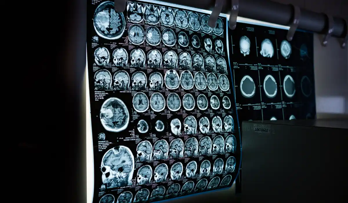
Digital subtraction angiography, or DSA, uses X-rays, contrast material, and computer subtraction to view blood vessels in the brain, neck, legs, lungs, or other body areas. DSA often uses a catheter and may help guide treatment.
The main difference is simple: fluorescein angiography focuses on the eye, while DSA checks blood vessels in other parts of the body.
How to Prepare for the Eye Angiogram?
Preparation for fluorescein angiography is usually simple. Your eye doctor may dilate your pupils before the test, so your vision may stay blurry and light-sensitive for several hours afterward.
Bring sunglasses for after the appointment. You may also want someone to drive you home, especially if your vision feels blurry after dilation. Tell your doctor about allergies, pregnancy, breastfeeding, kidney disease, and all medicines you take.
What to Tell Your Eye Doctor Before the Test?
Before fluorescein angiography, tell your eye doctor if you have ever had a reaction to fluorescein dye or any injected dye. Also mention asthma, severe allergies, heart disease, kidney problems, or any history of fainting during injections.
You should also tell your doctor if you are pregnant, may be pregnant, or are breastfeeding. This does not always mean you cannot have the test, but your doctor needs this information to decide the safest plan.
What Happens During the Procedure?
During the test, your pupils are usually dilated with eye drops. You will sit in front of a retinal camera, and the technician will take a few pictures before the dye injection.
Next, fluorescein dye is injected into a vein in your arm or hand. The camera then takes a series of pictures as the dye travels through the blood vessels in the retina. You may see flashes of light from the camera during the test.
Does a Fluorescein Angiogram Hurt?
Fluorescein angiography is not usually painful. You may feel a quick needle pinch when the dye is injected. Some people feel warm, flushed, slightly nauseated, or notice a brief unusual taste.
The camera does not touch the eye. However, the bright flashes may feel uncomfortable for some people. Most side effects are short-lived and improve soon after the test.
How Long Does the Fluorescein angiography Test Take?
The imaging part of fluorescein angiography is usually fairly quick. However, the full appointment may take longer because of check-in, pupil dilation, image setup, dye injection, and review.
Plan extra time for blurry vision after dilation. Your eyes may stay sensitive to light for several hours, so sunglasses can help when you leave the clinic.
What Happens After Fluorescein Angiography?
After fluorescein angiography, your skin may look slightly yellow for a short time. Your urine may also turn bright yellow or orange as your body clears the dye.
These color changes are expected and usually go away within a day or two. Your doctor may ask you to drink fluids unless you have been told to limit fluids for another medical reason.
Possible Side Effects and Risks
This eye imaging test is low risk for most people, but side effects can happen. Some people feel nausea, dizziness, dry mouth, sneezing, or a metallic taste. These symptoms usually pass quickly.
Rarely, a person may have an allergic reaction to fluorescein dye. Symptoms may include itching, hives, swelling, breathing trouble, or feeling faint. Tell the medical team right away if you feel unwell during or after the test.
Who May Need Extra Precautions?
Some people may need extra care before fluorescein angiography. This includes people with a history of dye reaction, severe allergies, asthma, kidney disease, pregnancy, or certain heart conditions.
Your eye doctor will compare the benefits and risks before ordering the test. In some cases, they may choose another imaging method or take extra precautions during the appointment.
What Fluorescein Angiography Results Can Show?
Fluorescein angiography results can show leaking blood vessels, blocked vessels, poor blood flow, swelling, abnormal new vessels, or scarring. These findings help the doctor understand what is happening inside the retina.
The results may guide treatment decisions. Depending on the condition, treatment may include observation, eye injections, laser treatment, medicine, or follow-up imaging.
When to Call a Doctor After the Eye Angiogram?
Call your doctor if you develop a rash, severe itching, swelling, trouble breathing, chest tightness, fainting, or worsening symptoms after fluorescein angiography. These may be signs of a reaction that needs medical attention.
You should also call if you have eye pain, sudden vision loss, or new severe vision changes. Mild blurry vision from dilation is common, but serious or worsening symptoms should be checked promptly.
Benefits of Fluorescein Angiography
Fluorescein angiography gives doctors detailed information about retinal blood flow. It can help detect disease activity that may not be obvious from a basic eye exam alone.
The test can also help track whether treatment is working. For people with diabetic retinopathy, macular edema, wet AMD, or retinal vein occlusion, this information can support more targeted eye care.
Final Thoughts
Fluorescein angiography is an important retinal imaging test that helps eye doctors examine blood vessels in the back of the eye. It can show leakage, blockage, abnormal vessel growth, and blood flow changes that may affect vision.
If your doctor recommends a fluorescein angiogram, ask why you need it, how to prepare, and what the results may mean. The test is usually quick, and the information it provides can help guide diagnosis, treatment, and follow-up care.
FAQs
Fluorescein angiography is an eye imaging test that uses fluorescein dye and a camera to photograph blood flow in retinal vessels.
Doctors use fluorescein angiography to check leaking, blocked, or abnormal retinal blood vessels linked to diabetic retinopathy, macular degeneration, or other eye diseases.
The test is usually not painful. You may feel a small needle pinch during dye injection and see bright camera flashes.
Fluorescein dye is safe for most people, but side effects can happen. Rarely, people may have an allergic reaction that needs medical care.
The photo-taking part is usually quick, but the full visit may take longer because of pupil dilation, preparation, dye injection, and follow-up instructions.
Your vision may stay blurry and light-sensitive after pupil dilation. Ask your clinic if you should bring someone to drive you home.