Medical imaging is an essential part of modern healthcare. Many people hear the terms sonogram and ultrasound interchangeably, but there are subtle differences worth understanding. Knowing the distinctions can help patients feel more informed when undergoing these imaging procedures.
Both methods use sound waves to create images of internal structures in the body. While the technology is largely similar, the terms reflect slightly different aspects of the process and output. Recognizing these differences can improve communication with healthcare providers and make the imaging experience less confusing.
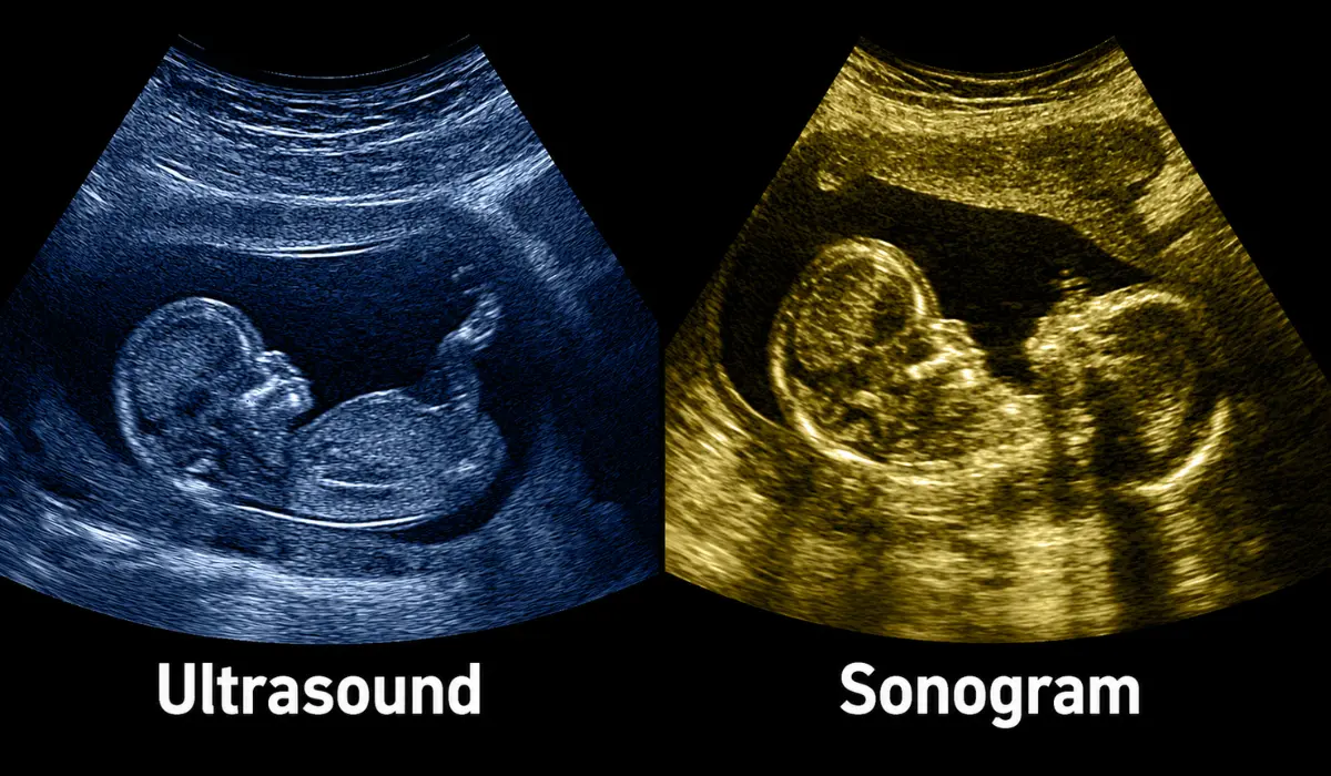
What Are Ultrasounds?
Ultrasound refers to the technology that uses high-frequency sound waves to capture images inside the body. This imaging technique is non-invasive, painless, and widely used in medical diagnostics.
Doctors often recommend ultrasound to monitor internal organs, check blood flow, or examine soft tissue structures. It is commonly associated with prenatal care, but its applications go far beyond pregnancy, including detecting abnormalities and guiding certain procedures.
Ultrasound machines generate sound waves that bounce off tissues and organs. These echoes are processed to create live images on a screen, allowing real-time observation of movement and function within the body.
What Are Sonograms?
A sonogram is the actual image or record created during an ultrasound exam. It is the visual result formed from sound waves and plays an important role in radiology imaging.
While ultrasound refers to the procedure and technology, a sonogram is the image that doctors and patients review after or during the scan. It can be printed, saved digitally, or used to show organs, soft tissues, blood flow, or a developing baby.
In radiology, healthcare providers study sonograms to detect abnormalities, monitor health conditions, and support treatment planning. These images help doctors explain findings clearly and give patients a better understanding of their medical care.
Key Differences Between Sonogram And Ultrasound
The main difference lies in usage. Ultrasound refers to the procedure and the technology, while sonogram refers to the image that results from the procedure. Think of it like taking a photograph: the camera is the ultrasound, the photo is the sonogram.
Understanding this distinction is helpful for patients reading medical reports. When a doctor mentions a sonogram, they are referring to the images captured during the ultrasound session, not the procedure itself.
Additionally, while ultrasound can describe real-time scanning, sonograms are static or captured snapshots. Modern machines may produce dynamic sonograms as well, showing video sequences of internal activity.
Common Uses Of Ultrasound And Sonograms
Ultrasound and sonogram technologies are widely used in healthcare. In obstetrics, sonograms monitor fetal growth and detect potential issues during pregnancy. The images allow parents and doctors to visualize development clearly.
Beyond prenatal care, ultrasounds and sonograms examine the abdomen, heart, blood vessels, and joints. They guide needle biopsies, detect gallstones, and assess organ health. Their safety and non-invasive nature make them preferable for many diagnostic applications.
Emergency medicine also relies on ultrasound for rapid assessment of internal injuries or fluid accumulation. Sonograms serve as documentation of these findings, providing a reliable reference for follow-ups.
Safety And Accessibility
Ultrasound procedures are generally safe, with no radiation exposure. This makes them suitable for repeated use and for vulnerable populations such as pregnant women and children.
Sonograms are easily accessible in hospitals, clinics, and imaging centers. The portability of some ultrasound machines allows for bedside or field assessments, increasing convenience and timely diagnosis.
Patients should feel comfortable asking healthcare providers about the purpose of each scan. Understanding what a sonogram will show can reduce anxiety and help in making informed healthcare decisions.
Interpreting Sonograms
Reading a sonogram requires medical training. Healthcare providers analyze shades, shapes, and movements to identify normal versus abnormal findings. They may compare multiple images to track changes over time.
For patients, sonograms provide visual confirmation of what is happening inside the body. Doctors often explain the images during or after the procedure to help patients understand their condition.
While sonograms are highly informative, interpretation should always be left to qualified professionals to avoid misunderstandings or misdiagnosis.
Benefits Of Ultrasound And Sonogram Imaging
These imaging methods are non-invasive, safe, and cost-effective. Ultrasounds provide real-time information about organ function, while sonograms offer tangible images for documentation and patient records.
The ability to visualize internal structures without surgery reduces risk and discomfort. Regular use can support early diagnosis, treatment planning, and ongoing health monitoring.
The clarity and accessibility of sonograms enhance communication between patients and healthcare providers, making medical care more transparent and reassuring.
Conclusion
Understanding the difference between sonogram vs ultrasound helps patients navigate medical imaging with more confidence. Ultrasound refers to the procedure and technology that uses sound waves to examine internal structures, while a sonogram is the actual image produced during that process.
Both are safe, non-invasive, and widely used in medical diagnostics, from monitoring pregnancy to assessing organs and blood flow. By knowing what each term means, patients can better interpret results, ask informed questions, and feel more engaged in their healthcare journey.
FAQs
They are related but not identical. Ultrasound refers to the procedure, while a sonogram is the resulting image produced by it.
Yes, ultrasound is generally safe, as it uses sound waves rather than radiation, making it suitable for monitoring fetal development.
Typically, an ultrasound takes 15-45 minutes depending on the area being examined and the complexity of the scan.
No, interpreting sonograms requires medical expertise. Healthcare providers are trained to read the images accurately and diagnose conditions.
Sonograms are painless. Patients may feel slight pressure from the transducer on the skin, but the process is non-invasive and comfortable.
References
RadiologyInfo.org – Ultrasound Imaging
https://www.radiologyinfo.org/en/ultrasound
Useful for explaining how ultrasound works and the resulting images (sonograms) used in medical diagnostics.
Mayo Clinic – Ultrasound Overview
https://www.mayoclinic.org/tests-procedures/ultrasound/about/pac-20395177
Helpful for understanding ultrasound procedures, common uses, and what sonogram images show.
Cleveland Clinic – Sonogram and Ultrasound
https://health.clevelandclinic.org/sonogram-vs-ultrasound
Provides details about the difference between the ultrasound procedure and the sonogram image.
Johns Hopkins Medicine – Ultrasound Imaging
https://www.hopkinsmedicine.org/imaging/exams-and-procedures/ultrasound
Good source for patient-friendly explanation of ultrasound technology and how sonograms are produced.