If your doctor has recommended a CT scan, you may have questions about what it is, why it’s needed, and what the procedure involves. While the name may sound intimidating, a CT scan is one of the most common and valuable diagnostic tools used in modern healthcare.
Every day, millions of patients undergo CT scans to help doctors identify injuries, diagnose illnesses, and monitor treatment progress.
From emergency rooms to outpatient imaging centers, CT technology plays a critical role in helping healthcare providers see inside the body without surgery.
Understanding how a CT scan works can help patients feel more comfortable and informed before their appointment.
What Is A CT Scan?
A CT scan, short for Computed Tomography scan, is a medical imaging test that uses X-rays and computer technology to create detailed pictures of the inside of the body.
Unlike a traditional X-ray that produces a single image, a CT scan captures multiple images from different angles. A computer then combines these images to create detailed cross-sectional views of bones, organs, blood vessels, and soft tissues.
These images allow doctors to examine areas of the body with much greater detail than standard X-rays can provide.
How Does A CT Scan Work?
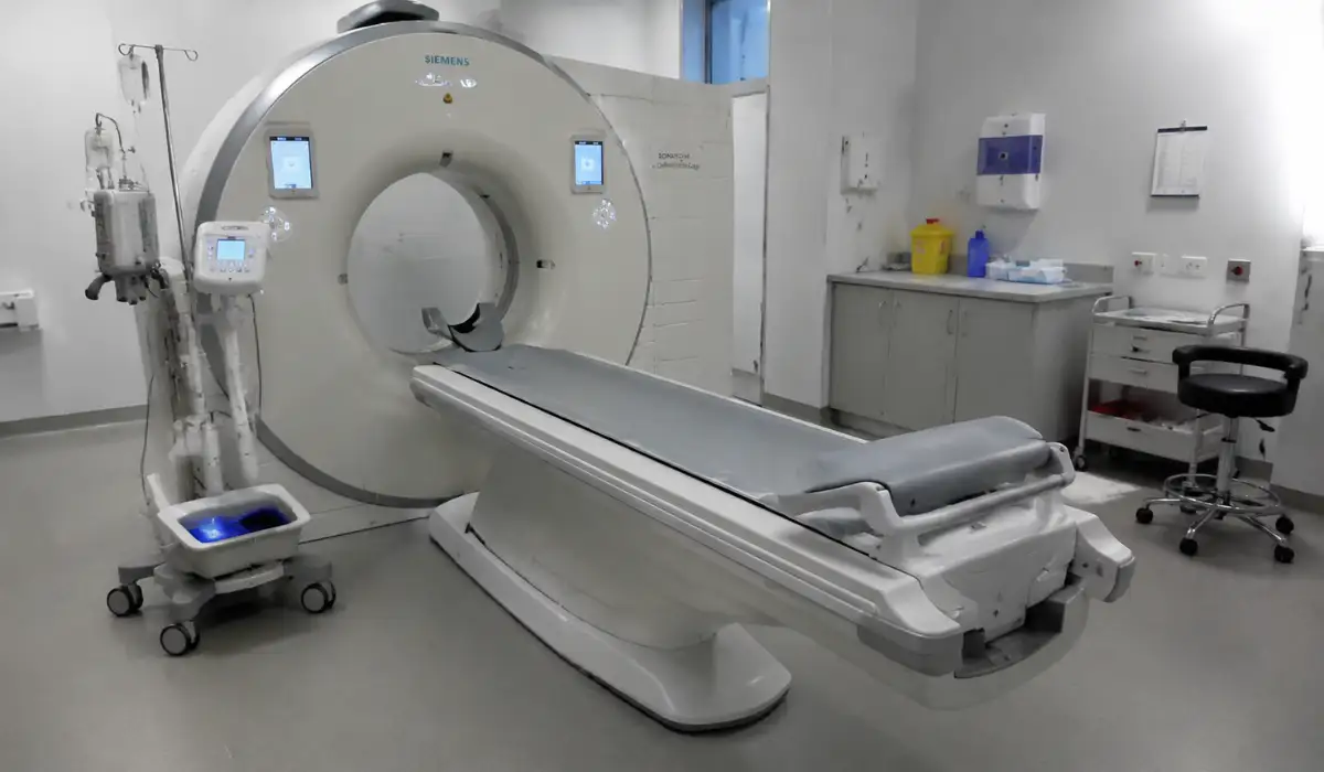
A CT scanner is a large, donut-shaped machine with an opening in the center. During the exam, the patient lies on a motorized table that slowly moves through the scanner.
As the table moves, an X-ray tube rotates around the body, capturing images from multiple angles. Advanced computer software processes these images and creates detailed slices of the area being examined.
The procedure is painless, and most scans take only a few minutes to complete.
Why Do Doctors Order CT Scans?
CT scans help doctors investigate a wide range of symptoms and medical conditions. Because they provide detailed images quickly, they are often used when an accurate diagnosis is needed.
Common reasons for a CT scan include:
- Unexplained pain
- Head injuries
- Suspected strokes
- Bone fractures
- Internal bleeding
- Tumors or cancer evaluation
- Lung conditions
- Heart and blood vessel problems
- Kidney stones
- Abdominal disorders
In emergency situations, CT scans often provide critical information that helps physicians make rapid treatment decisions.
What Parts Of The Body Can Be Scanned?
CT scans can examine nearly any part of the body.
Head and Brain
Doctors use CT scans to evaluate strokes, brain injuries, bleeding, tumors, and neurological conditions.
Chest
Chest CT scans help assess the lungs, heart, and blood vessels. They are commonly used to investigate infections, chest pain, and respiratory conditions.
Abdomen and Pelvis
These scans can help diagnose digestive disorders, kidney stones, liver disease, appendicitis, and other abdominal conditions.
Bones and Joints
CT imaging provides detailed views of fractures, joint damage, and orthopedic injuries.
Blood Vessels
Specialized CT angiography exams allow doctors to evaluate blood flow and detect blockages or abnormalities within blood vessels.
What Is Contrast Material?
Some CT scans require the use of contrast material, often called contrast dye.
Contrast helps certain tissues, organs, and blood vessels appear more clearly on the images. Depending on the examination, contrast may be:
- Injected into a vein
- Swallowed as a liquid
- Administered through other methods
Not every CT scan requires contrast. Your healthcare provider will determine whether it is necessary for your specific exam.
What Should Patients Expect During The Procedure?
Most CT scans are straightforward and comfortable.
Typically, patients will:
- Change into a gown if necessary.
- Remove metal objects such as jewelry.
- Lie on the scanning table.
- Remain still while images are taken.
- Follow breathing instructions when requested.
The machine may produce soft buzzing or clicking sounds, but the scan itself is painless.
Most patients are able to resume normal activities immediately after the procedure.
Benefits Of CT Scans
CT scans offer several advantages that make them one of the most frequently used imaging tests.
Detailed Imaging
CT technology provides highly detailed images of bones, organs, blood vessels, and soft tissues.
Fast Results
Many scans can be completed within minutes, making them especially valuable during emergencies.
Non-Invasive
The procedure does not require surgery or major intervention.
Accurate Diagnosis
The detailed images help healthcare providers identify conditions that might not be visible through physical examination alone.
Treatment Planning
CT scans help physicians determine the best treatment options and monitor patient progress over time.
Are CT Scans Safe?
CT scans are generally considered safe and are performed millions of times each year.
Because CT imaging uses X-rays, patients are exposed to a small amount of radiation. However, healthcare providers carefully consider the benefits and risks before recommending any imaging study.
For most patients, the value of obtaining an accurate diagnosis far outweighs the minimal risks associated with radiation exposure.
Patients should inform their healthcare provider if they are pregnant or think they may be pregnant before undergoing a CT scan.
How Are CT Scans Different From MRI Scans?
Although both CT scans & MRI scans create detailed images of the body, they use different technologies.
CT scans use X-rays and are typically faster. They are often preferred for emergency situations, trauma cases, and bone imaging.
MRI scans use magnetic fields and radio waves. They are often better suited for evaluating soft tissues such as the brain, spinal cord, ligaments, and muscles.
Your healthcare provider will choose the imaging test that best fits your medical needs.
Conclusion
A CT scan is one of the most important diagnostic tools in modern medicine. By combining X-ray technology with advanced computer processing, CT scans provide highly detailed images that help doctors diagnose injuries, detect diseases, and guide treatment decisions.
Whether evaluating abdominal pain, investigating a head injury, monitoring cancer treatment, or diagnosing a lung condition, CT scans provide valuable information that supports better patient care.
Understanding how the procedure works can help patients feel more confident and prepared when undergoing this common medical imaging test.
Frequently Asked Questions
A CT scan stands for Computed Tomography scan. It’s an imaging method that combines X-rays with computer processing to create detailed cross-sectional images of the body.
Unlike a standard X-ray that produces a flat, 2D image, a CT scan captures multiple angles and creates 3D images. This allows doctors to see organs, bones, and tissues in more detail.
CT scans are generally safe when performed under medical guidance. They involve low levels of radiation, but doctors weigh the benefits versus risks before recommending a scan.
Preparation depends on the type of CT scan. Sometimes fasting is required, or a contrast dye may be used to highlight blood vessels and organs. Your healthcare provider will provide specific instructions.
Most CT scans are quick, usually taking between 10-30 minutes, including setup time. The actual scanning part often takes just a few minutes.
CT scans are excellent for detecting many conditions, including bone fractures, tumors, blood clots, and internal injuries. However, some conditions may require other imaging methods like MRI or ultrasound.
The CT scan itself is painless. You may feel slight discomfort from lying still on the table or from the contrast dye injection if used.
Typically, a radiologist reviews the images and sends a report to your doctor within 24-48 hours, though urgent cases may be reviewed sooner.