The inferior rectus muscle is one of the six extraocular muscles that move the eye. Its main job is to help move the eye downward, especially when looking down to read, walk downstairs, or shift gaze toward the lower field of vision.
This eye muscle also helps with adduction and outward rotation of the eyeball. Understanding the inferior rectus muscle matters because weakness, restriction, trauma, thyroid eye disease, or nerve problems can cause double vision, eye misalignment, and difficulty looking down.
What to Know About the Inferior Rectus Muscle?
The inferior rectus muscle is controlled by the oculomotor nerve, also called cranial nerve III. This nerve helps several eye muscles coordinate normal eye movement.
Its primary function is eye depression, but it also contributes to adduction and extorsion. These movements help the eyes track objects while reading, walking, or looking downward.
Inferior rectus restriction may occur in thyroid eye disease or orbital floor fracture. These conditions can make the eye feel stuck or cause vertical double vision.
Inferior rectus palsy is different from muscle tightness. Palsy means weakness from nerve or muscle dysfunction, while restriction means the eye muscle cannot move freely.
Sudden double vision, eye pain, bulging eye, trauma, or drooping eyelid needs medical evaluation. These symptoms may involve the eye muscles, cranial nerves, orbit, or brain.
Overview Table
| Feature | Inferior Rectus Muscle Details |
| Muscle group | Extraocular muscle |
| Location | Lower part of the orbit beneath the eyeball |
| Main action | Moves the eye downward |
| Other actions | Helps move the eye inward and rotate it outward |
| Nerve supply | Oculomotor nerve, inferior division |
| Works with | Superior rectus, superior oblique, inferior oblique, medial rectus, lateral rectus |
| Common symptom when affected | Double vision or vertical eye misalignment |
| Common related conditions | Strabismus, thyroid eye disease, orbital fracture, cranial nerve III palsy |
| Common tests | Eye movement exam, cover test, prism test, CT, MRI, thyroid blood tests |
| Specialist | Ophthalmologist, neuro-ophthalmologist, pediatric ophthalmologist, or strabismus specialist |
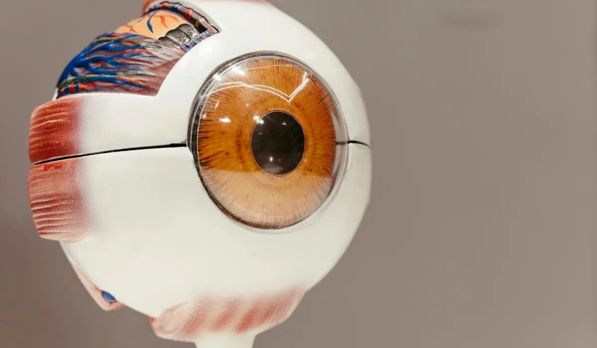
Inferior Rectus Muscle Anatomy and Function
The inferior rectus muscle is part of the extraocular muscle system, which controls how the eyes move. These muscles are outside the eyeball but inside the eye socket, also called the orbit.
The inferior rectus starts near the back of the orbit from a fibrous structure called the common tendinous ring. It then runs forward under the eyeball and attaches to the lower front surface of the eye.
When this lower rectus muscle contracts, it pulls the eye downward. This is why it plays an important role in looking down while reading, checking steps, using a phone, or focusing on something below eye level.
Common Causes of Inferior Rectus Muscle Issues
People often search for inferior rectus muscle anatomy because they have double vision, abnormal eye movement, thyroid eye disease, or an orbital fracture report mentioning the muscle. The term may also appear in MRI, CT scan, or ophthalmology notes.
This muscle is clinically important because the eyes must move together. Even a small difference in movement between both eyes can create diplopia, eye strain, headaches, or trouble judging depth.
The inferior rectus can be affected by nerve palsy, inflammation, trauma, scarring, surgery, or thyroid-related eye muscle enlargement. Each cause has a different treatment approach, so a proper eye examination is important.
Inferior Rectus Muscle Symptoms and Warning Signs
Inferior rectus muscle problems may cause difficulty looking down, especially when the affected eye does not move normally. Some people notice double vision when reading, walking downstairs, or looking toward the lower side.
Vertical strabismus may occur when one eye sits higher or lower than the other. This can make images appear stacked on top of each other rather than side by side.
Other possible signs include eye pain, eye pressure, abnormal head tilt, eye fatigue, bulging eyes, eyelid drooping, or limited eye movement. Symptoms can vary depending on whether the issue is muscle weakness, tightness, entrapment, or nerve involvement.
How Inferior Rectus Muscle Problems Are Diagnosed?
A doctor evaluates inferior rectus function by checking eye alignment, eye movement, and whether double vision changes in different gaze positions. The exam may include a cover test, prism measurements, pupil exam, and visual acuity testing.
An ophthalmologist may ask the patient to follow a target up, down, left, and right. Limited downward movement, vertical diplopia, or abnormal eye position can help identify whether the inferior rectus muscle, another extraocular muscle, or a cranial nerve is involved.
Imaging may be needed when trauma, thyroid eye disease, orbital mass, inflammation, or nerve disease is suspected. CT scans are commonly used for orbital fracture, while MRI may help assess soft tissue, nerves, and brain-related causes of eye movement problems.
Inferior Rectus Muscle Treatment Options
Treatment for an inferior rectus muscle problem depends on the cause. There is no single treatment for every case of double vision, strabismus, or restricted downward gaze.
Temporary options may include prism glasses, patching one eye, lubricating drops, or monitoring while inflammation improves. These approaches may reduce symptoms but do not treat every underlying cause.
Some cases require thyroid disease treatment, anti-inflammatory therapy, eye muscle surgery, orbital fracture repair, or management of a cranial nerve problem. A strabismus specialist or neuro-ophthalmologist can decide which option fits the diagnosis.
Practical Steps Readers Can Follow
Anyone with new double vision, eye movement restriction, or a CT scan mentioning the inferior rectus muscle should arrange an eye examination. Bring imaging reports, medication lists, thyroid history, and details about when symptoms started.
Avoid driving if double vision affects safety. Covering one eye may temporarily reduce binocular diplopia, but this should not replace medical evaluation.
After an eye injury, avoid pressing on the eye or blowing the nose forcefully until a clinician checks for orbital fracture. Trauma-related inferior rectus entrapment can need urgent assessment, especially when nausea, severe pain, or restricted eye movement is present.
Inferior Rectus Muscle Myths and Facts
One misconception is that all double vision comes from the inferior rectus muscle. Diplopia can also come from cornea problems, cataracts, cranial nerve palsy, thyroid eye disease, stroke, myasthenia gravis, or other eye muscle disorders.
Another misconception is that eye exercises can fix every eye muscle problem. Exercises may help selected conditions, but they cannot release an entrapped inferior rectus muscle or correct all forms of strabismus.
Some people assume a normal vision chart means the eye muscles are healthy. Visual acuity can be normal even when extraocular muscle movement or eye alignment is abnormal.
A further misconception is that thyroid eye disease only affects thyroid hormone levels. In reality, thyroid-related eye disease can affect orbital tissues and extraocular muscles, including the inferior rectus.
Inferior Rectus Muscle Risks and Complications
Untreated inferior rectus restriction or palsy may lead to persistent double vision, abnormal head posture, eye strain, reduced depth perception, and difficulty with reading or stairs. In children, untreated eye misalignment may affect visual development.
Orbital trauma can cause swelling, bleeding, fracture, or muscle entrapment. If the inferior rectus is trapped in an orbital floor fracture, eye movement may become restricted and symptoms may worsen without proper care.
Eye muscle surgery can improve alignment in selected cases, but it carries possible risks such as overcorrection, undercorrection, scarring, infection, or persistent diplopia. The decision should be made with an experienced eye specialist.
When to Seek Professional Help?
Seek prompt medical care for sudden double vision, new eye misalignment, eye pain, drooping eyelid, enlarged pupil, severe headache, weakness, numbness, or trouble speaking. These symptoms may involve the cranial nerves or brain.
After facial or eye trauma, urgent care is needed if there is double vision, restricted eye movement, nausea, vomiting, cheek numbness, sunken eye appearance, or severe swelling. These signs may suggest orbital fracture or inferior rectus entrapment.
Children with persistent eye turning, head tilt, or abnormal tracking should have a complete eye exam. Early assessment can help detect strabismus, amblyopia risk, or congenital eye muscle problems.
Questions to Ask a Professional
Ask whether the problem is caused by inferior rectus weakness, muscle restriction, nerve palsy, thyroid eye disease, or orbital trauma. These causes can look similar but require different treatment plans.
Useful questions include:
- Is the problem caused by inferior rectus weakness, muscle restriction, nerve palsy, thyroid eye disease, or orbital trauma?
- Which eye muscle is affected?
- Could another extraocular muscle also be involved?
- Is the oculomotor nerve or another cranial nerve causing the eye movement problem?
- Do I need CT or MRI imaging to check the orbit, nerves, or eye muscles?
- Would prism glasses help manage double vision?
- Is eye muscle surgery needed?
- Is monitoring more appropriate than immediate treatment?
- Are the extraocular muscles enlarged because of thyroid eye disease?
- Is thyroid eye disease active or inactive?
- How does thyroid eye disease affect treatment timing or surgical planning?
Patients with thyroid disease should ask whether the extraocular muscles are enlarged and whether thyroid eye disease is active or inactive. This can affect timing of treatment and surgical planning.
Conclusion
The inferior rectus muscle helps move the eye downward and supports coordinated eye movement with the other extraocular muscles. Problems with this muscle may cause double vision, vertical strabismus, restricted gaze, or difficulty looking down.
The most important step is identifying the cause. Inferior rectus muscle weakness, restriction, entrapment, thyroid eye disease, and nerve problems need different evaluation and treatment approaches.
FAQS
The inferior rectus muscle mainly moves the eye downward. It also helps move the eye inward and rotate the top of the eye outward during coordinated extraocular movement.
The inferior division of the oculomotor nerve, also called cranial nerve III, controls the lower rectus eye muscle. This nerve also supports several other important eye movements.
Yes. Weakness, tightness, swelling, or entrapment of this eye muscle can make the eyes point in different directions, causing vertical double vision or gaze-related diplopia.
Doctors check eye movement, alignment, prism measurements, and gaze-related symptoms. CT or MRI may be used when orbital fracture, thyroid eye disease, inflammation, or mass effect is suspected.
No. Inferior rectus palsy means the muscle or its nerve supply is weak. Strabismus means the eyes are misaligned, which may result from palsy, restriction, or other causes.
Thyroid eye disease can enlarge or stiffen extraocular muscles. The inferior rectus is commonly involved, which may restrict upward movement and contribute to vertical double vision.
Yes. An orbital floor fracture can trap or restrict tissues near the lower eye muscle. This may cause pain, limited eye movement, nausea, and double vision after facial trauma.
Eye exercises may help certain coordination problems, but they do not treat every inferior rectus disorder. Entrapment, thyroid-related restriction, or nerve palsy requires professional diagnosis.
An ophthalmologist usually evaluates extraocular muscle problems. A neuro-ophthalmologist, pediatric ophthalmologist, orbital specialist, or strabismus surgeon may be needed for complex cases.
Double vision with sudden headache, weakness, numbness, eye injury, severe pain, drooping eyelid, enlarged pupil, or speech trouble needs urgent medical care. These symptoms may signal serious disease.